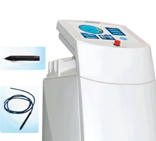
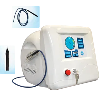
Facial telangiectasia treatment comes down to one idea: heat the blood inside a dilated vessel until its wall coagulates, then let the body clear it, without cooking the skin above it. Two routes work on the face. High frequency thermocoagulation through a fine contact tip, and vascular lasers such as long pulse 1064 nm Nd:YAG. Here's how to choose, and what to check before you sign a purchase order.
Why do these vessels show up on the face?
Telangiectasia appears when a capillary that should be invisible widens enough to show through the skin. Our technical archive puts it plainly: in simple telangiectasia the capillaries of the dermal papilla dilate to roughly 0.1 to 1.0 mm. Hence the fine red threads across cheeks, nose and chin, rather than a flat blush.
The triggers stack up over years. Alcohol, oestrogen, corticosteroids and chronic photoaging all appear in our dermatology training material as recognised contributors, with a note that these vessels favour lighter Fitzpatrick skin types and that rosacea travels with them.
Diagnosis matters more than the machine. Threads on a cheek are not a congenital nevus flammeus or a raised angioma, and won't answer to the same settings. This guide stays with the discrete vessels grouped under single telangiectasis.
What selective targeting actually does in tissue
Every light-based vascular treatment traces back to selective photothermolysis, published by Anderson and Parrish in Science in 1983. Pick a wavelength the target absorbs far more strongly than its neighbours, deliver it in a pulse short enough that heat stays put, and the vessel takes the damage instead of the dermis.
Hemoglobin does the absorbing. Our technical archive lists oxyhemoglobin absorption peaks at 418 nm, 542 nm and 577 nm, which is why pulsed dye lasers were built around the yellow end of the spectrum. A second effect matters: heated hemoglobin oxidises toward methemoglobin, which absorbs 1064 nm far more strongly. That's part of why a long pulse Nd:YAG keeps working on a vessel it has begun to heat.
High frequency reaches the same endpoint without light. A fine insulated tip touches the vessel and drives radiofrequency current into it until the hemoglobin and the endothelial cells of the wall coagulate and the capillary seals. Our device manuals for the high frequency vascular platforms record a 13.56 MHz output, working frequency adjustable from 1000 to 2000 Hz in 100 Hz steps, continuous and pulsed modes, and a treatment tip about 0.01 mm across. Coagulated tissue sloughs off in three to seven days. The physics is in our explainer on selective photothermolysis.
High frequency or vascular laser: which one for facial vessels?
Neither wins outright. High frequency shines on discrete surface threads you can trace with a tip. Lasers and IPL cover diffuse redness and deeper vessels far faster. A clinic seeing rosacea patients and one-off spider veins usually owns both.
| Factor | High frequency | Vascular laser / IPL |
|---|---|---|
| Energy source | Radiofrequency current, fine contact tip | Light absorbed by hemoglobin |
| Best target | Discrete threads, spider veins, cherry angiomas | Diffuse redness, broad fields, deeper vessels |
| Melanin competition | None, no wavelength involved | Real below 800 nm; longer wavelengths safer on dark skin |
| Main risk if pushed | Crusting and marking at the tip | Burns and pigment change from poor cooling |
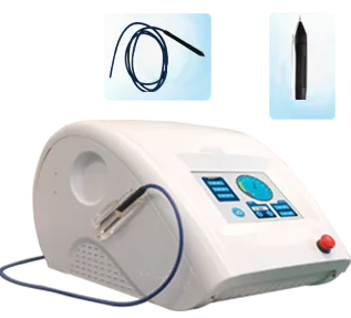
Our own range sits behind that first column. The high frequency vascular platforms we build share the 13.56 MHz output described above, the adjustable 1000 to 2000 Hz working frequency, the 0.01 mm treatment tip, and both continuous and pulse modes. Individual units differ in console and handpiece configuration, so ask for the current specification sheet against the model you're quoting rather than working from a number in an old catalogue.
On the laser side, the standard guidelines of care for vascular lesions in the Indian Journal of Dermatology, Venereology and Leprology (Srinivas and Kumaresan, 2011) put pulsed dye laser first on efficacy and safety evidence, with Nd:YAG and IPL as the other common options.
Want the parameter sheet before you shortlist?
Send us the lesions you see most, your usual patient skin types and your weekly volume. We'll return parameter sheets for the matching Pmise models and a straight technical view on whether high frequency, a vascular laser, or both belongs in your room. Use the Pmise enquiry form, or browse the high frequency vascular range.
Where long pulse Nd:YAG earns its place
Reach for 1064 nm when the vessel is too deep, too blue or too broad for a tip. Our device manuals describe the long pulse 1064 nm beam as selectively absorbed by hemoglobin and able to penetrate the epidermis to the target vessel, with adjustable spot size and sapphire contact cooling.
Why not 532 nm? Our dermatology training material is blunt: green light scatters heavily at the epidermis and risks pigmentary change, so 1064 nm is the recommended choice for dilated facial vessels. The spot runs small, roughly 3 to 6 mm. Precise, but slow across a large field. Our piece on vascular conditions and Nd:YAG covers parameters.
Cooling is where facial vascular work goes wrong
Here's the counterintuitive part. More cooling is not automatically safer. Our technical archive gives a rule: when the target sits in the dermal papilla, immediate epidermal cooling is not recommended, because chilling the surface during the pulse also drains heat from a target a fraction of a millimetre below. Cool actively before and after the pass instead.
Deeper lesions flip the logic. Port wine stains and larger malformations need high fluence, and that needs robust epidermal cooling or you get blistering. Match cooling to depth, not habit.
A conservative facial protocol
One thing before the steps. This is a device operating reference for trained practitioners, not medical advice and not a diagnosis. Vascular lesion treatment should be performed by a qualified medical or aesthetic professional, within the scope their own jurisdiction permits. Device registration status and approved indications differ from market to market, so verify local regulations and the licensing position of any model before you buy it or treat with it.
Facial skin is unforgiving. Restraint beats power.
- Take a real history: photosensitising medication, recent tanning, isotretinoin, pregnancy, bleeding disorders, keloid tendency, prior hyperpigmentation.
- Confirm the lesion. Simple telangiectasia, rosacea erythema, angiomas and malformations behave differently under one handpiece.
- Clean the skin properly. Make-up and sunscreen residue compete for absorbed energy.
- Test first. Small area, conservative settings, watch the endpoint, then decide whether to step up. Never mid-pass, on a hunch.
- Set cooling to depth. Light or no immediate cooling for superficial threads; contact cooling for deeper laser work.
- Brief the patient before they leave the chair: no picking at crusts, daily SPF, no heat or alcohol for a few days.
Check skin type against your settings, not a factory preset. Our guide to Fitzpatrick skin types and laser settings covers types IV and up.
What buyers should check before ordering a vascular platform
Clinical fit is half the decision. What happens after the crate is opened decides the rest. Ask for each item below in writing, against the model and your market.
- Conformity for your market. Ask which assessment route the model went through, request certificate copies, and check their scope covers the device you're buying. Our comparison of medical CE versus standard CE and the piece on the US market route show what to look at.
- Warranty and spare parts. Get the warranty period, its exclusions, and how long critical parts stay available. Ask which ship from stock and which are built to order.
- Training and parameter support. Who trains your operators, in what language, and does parameter guidance come with the machine or cost extra? A vascular device in untrained hands marks faces. That matters more than sticker price.
- Consumables and handpiece life. Fine treatment tips, handpieces and optical parts wear out. Ask for the replacement list with unit prices and the reorder process after year one.
- Lead time and shipping terms. Confirm production lead time, the Incoterm on the quotation, packing dimensions, and voltage and plug configuration.
- Distribution and OEM terms. Pin down territory, order commitments, demo unit support, and whether branding and interface language can be adapted.
Two guides go deeper: warranty and spare parts and running costs and consumables.
What results should you promise?
Be specific and be modest. According to the American Academy of Dermatology, most patients treated with laser or light for the visible blood vessels of rosacea see a 50% to 75% reduction after one to three treatments, spaced three to four weeks apart. New vessels can still form, the AAD notes, since the condition is chronic.
Treat that as a reference point, not a promise. Outcomes shift with vessel size, depth, skin type and operator technique, and no device should be sold or consented on a guaranteed result. Redness afterwards is normal, and the AAD describes it as usually fading within two weeks.
Clinics getting consistently good vascular results aren't running the highest fluence. They diagnose carefully and test first.
Frequently Asked Questions
Is high frequency or laser better for facial spider veins?
It depends on the vessel, not the brochure. A fine-tip high frequency device suits discrete surface threads you can trace individually, and many close in one pass. Lasers and IPL are better for diffuse redness across a cheek, or deeper vessels. Most busy clinics keep both.
How many sessions does facial telangiectasia treatment take?
Isolated vessels treated point by point often need very few passes. For rosacea-associated redness, the American Academy of Dermatology reports most patients see a 50% to 75% reduction after one to three laser or light treatments, three to four weeks apart. Plan a short course rather than promising clearance.
Does treating facial vessels hurt or leave marks?
Most patients report brief stinging rather than real pain, and pulsed output modes exist partly to improve comfort. Temporary redness is expected. With high frequency work, tiny crusts form over treated vessels and separate in roughly three to seven days according to our device manuals. Scarring is uncommon at conservative energy.
Will the veins come back after treatment?
A vessel that has been properly coagulated is cleared by the body, and the AAD states treated vessels don't reappear. What comes back is the tendency. Rosacea stays chronic, sun damage accumulates, and new telangiectasia can appear later. Sun protection and rosacea management stretch the result.
Next step. Specifying a vascular room, or adding the category to a distribution portfolio? Send your case mix, skin types and destination market. Back comes the parameter sheet, plus written answers on training, warranty and spare parts. Start at the Pmise enquiry form or the high frequency vascular line.
Pmise Technical Team. Pmise (pameisi.com) builds laser, light and high frequency aesthetic systems and supports clinics and distributors worldwide on device selection, parameters and operator training.