
Pigment Treatment
Blue Nevus Treatment: 1064nm Q-Switched Nd:YAG Protocol and Clinical Guide
A blue nevus is a small blue-grey mole built from pigment cells that sit deep in the dermis rather than on the surface. Most never need touching. When one is addressed for cosmetic reasons, blue nevus treatment means either surgical excision or a 1064nm Q-switched Nd:YAG laser, picked because that wavelength reaches deep dermal pigment.
This page is educational material for clinics, dermatologists, and distributors evaluating equipment. It isn't medical advice, and no lesion should be treated without an in-person assessment.
What a blue nevus actually is
Blue nevi are benign melanocytic lesions. Under the microscope you see spindle-shaped or ovoid dendritic melanocytes with heavily pigmented cytoplasm, sitting wholly or partly within the dermis. Our engineering archive says the same thing in plainer words: dermal melanocytes, deep in the dermis, packed with melanin granules.
So why blue? The pigment is brown. Light scattering in the skin sends shorter blue wavelengths back out from that depth, so a deep brown deposit reads as blue-grey. Blue means deep, and depth drives every decision that follows.
Two presentations are worth separating:
- Common blue nevus. A solitary grey-blue macule, papule or small nodule with a sharp border, growing slowly. DermNet puts the usual size at 0.5 to 1 cm; our internal skin-lesion reference records 1 to 8 mm. Face and arms are typical sites, along with the backs of the hands and feet.
- Cellular blue nevus. Larger, more nodular, blue to blue-black, and usually at least 1 cm across. It favours the buttock and sacrococcygeal region and is often present from birth. This is the variant linked with malignant transformation, and it changes the whole conversation.
Combined, sclerosing and epithelioid forms exist too. For equipment decisions, the split above is what matters.
Diagnosis comes first, and it isn't optional
Here is the uncomfortable part for anyone selling or operating a laser. Standard dermatology guidance says a blue nevus usually needs no treatment at all. DermNet is direct: lesions larger than 1 cm, lesions that change, and lesions that appear new in an adult should be considered for histological evaluation to exclude melanoma. Our own reference material lands in the same place. Leave stable lesions under 1 cm alone. Excise the larger ones promptly.
A laser destroys exactly the tissue a pathologist needs to read. Fire first and you may erase evidence of an early malignancy while leaving the deep cells behind. Blue nevi also sit deep enough that a shave biopsy can return an incomplete picture, so excisional sampling is generally preferred.
Practical rule for the treatment room: no histology, no laser. If the lesion is new in an adult, growing, changing colour, bleeding, or over 1 cm, it goes to a dermatologist or surgeon, not onto the handpiece.
How a Q-switched 1064 laser targets dermal melanocytes
The mechanism is selective photothermolysis, described by Anderson and Parrish in Science in 1983. Deliver a pulse shorter than the target's thermal relaxation time, at a wavelength the target absorbs strongly, and the energy stays inside the target. Melanosomes fracture in nanoseconds. Surrounding collagen and vessels barely notice.
Wavelength choice is about reach. Our device manuals put it simply: 1064nm removes blue, black and green-blue pigment, while 532nm handles red and superficial colour. Longer wavelengths scatter less on the way down, so 1064nm still has useful energy left at dermal depth. That is why it dominates Nevus of Ota, Hori nevus, and dark tattoo work.
Spot size matters more than most operators expect. A wider beam loses proportionally less energy to lateral scattering, so it reaches deeper at the same fluence. Our manuals advise a spot above 2 mm for dermal pigment, reserving smaller spots for junctional lesions and scar work. Go small and hot on a deep target and you concentrate injury in the epidermis.
One honest caveat. A blue nevus is a nevus-cell tumour, not the loose scatter of pigment-laden cells in Nevus of Ota. Clearance is slower and less predictable, partial fading is common, and recurrence happens. Anyone promising a clean result in two sessions is guessing.
Treatment protocol and what to expect
These starting points come from our device manuals for dermal melanocytic lesions, the closest documented analogue. Treat them as a test-patch starting line, not a prescription. Titrate up from a low setting.
| Parameter | Documented starting point (dermal pigment) | Notes |
|---|---|---|
| Wavelength | 1064nm | 532nm suits superficial and red pigment, not this lesion |
| Spot diameter | 3 mm and above | Larger spots reach deeper |
| Repetition rate | 3 to 10 Hz | Set by operator skill, not ambition |
| Single pulse energy | From about 400 mJ, titrated up | Under 200 mJ is unsuitable for dermal work |
| Clinical endpoint | Mild immediate whitening of the treated spot, fading within minutes | Stop the pass at that point |
| Signs the fluence is too high | Pinpoint bleeding, pronounced purpura, tissue splatter, grey blanching spreading past the lesion margin | Drop the energy. Over-treatment is what scars |
| Interval | Roughly 8 to 12 weeks | Rushing raises PIH risk |
| Session count | Several, often many | Partial clearance is a realistic goal |
Watch the skin, not the display. Published nanosecond Q-switched protocols for dermal melanocytic pigment define the therapeutic endpoint as immediate whitening after the pulse, and the same literature reports reducing energy density when tissue bleeding becomes prominent. Faint frost appears, you move on. Blood appears, you back off.
Recovery follows a predictable arc. The whitening settles within minutes, then mild swelling arrives, then a light scab. Let the crust separate on its own. Visible lightening usually starts around a month out, and the lesion may darken for two to four weeks first, because fragmented pigment migrates upward before it clears.
Set expectations in writing before the first pass. Results vary with lesion type, depth, size, and Fitzpatrick skin type. Darker phototypes carry a real risk of post-inflammatory hyperpigmentation, which is why conservative fluences and long intervals beat aggressive passes. For the pigment physics across lesion types, see our guide to treating pigmented lesions with a Q-switched laser, then compare this protocol against Nevus of Ota treatment, where the same wavelength behaves far more predictably.
Recommended Pmise equipment
Deep dermal pigment punishes underpowered machines. If single pulse energy is too low to fragment melanosomes, you don't get a slower result, you get no result plus repeated futile sessions. Our internal manuals are blunt: under roughly 200 mJ per pulse is unsuitable for dermal lesions.
For this category we point clinics to the Q-switched Nd:YAG laser range. Documented specifications from our manuals include:
- Dual wavelength output at 1064nm and 532nm from a single Nd:YAG cavity
- Flat-top beam profile, spreading energy evenly instead of peaking in the centre
- Spot diameter adjustable from 1 to 7 mm
- Single pulse energy up to 850 mJ at 1064nm and up to 600 mJ at 532nm
- Articulated arm delivery for steady handling through long sessions
The Pmise QN series covers general Q-switched pigment and tattoo work. The Pmise QE series uses electro-optic Q-switching, giving continuously adjustable pulse energy rather than fixed steps, and that granularity earns its keep when you're creeping up on a whitening endpoint in a deep lesion on darker skin. Clinics also handling raised lesions after histology usually pair a Q-switched platform with a CO2 device from the Pmise CF series, under local anaesthesia. Ask us for parameter sheets before committing.
Aftercare and precautions
- Cooling or barrier gel right after the pass, then keep the site moist
- Leave scabs alone. Picking is the fastest route to a scar
- Strict sun protection until pigment settles, ideally for several months
- Use a bland repair serum once the crust separates naturally
- Wavelength-rated eyewear for everyone in the room, patient included
- Test patch first on any Fitzpatrick IV to VI patient, then wait a full interval
- Photograph the lesion with a scale at every visit. If it changes shape, stop and refer
- File the histology report before session one, not after
Complications in trained hands are uncommon but real: post-inflammatory hyperpigmentation, transient hypopigmentation, persistent erythema, occasionally scarring. Patients with a keloid history are poor candidates. So are patients who can't commit to sun avoidance.
Frequently Asked Questions
Can a laser remove a blue nevus completely?
Sometimes, but don't promise it. The pigment cells sit deep and cluster densely, so response ranges from significant fading to stubborn persistence. Partial clearance over multiple sessions is the realistic framing. Where complete removal is the goal, particularly above 1 cm, surgical excision stays the definitive option and produces a specimen for histology.
Why 1064nm rather than 532nm or a picosecond device?
1064nm scatters least on the way down, so it delivers usable energy at the depth this lesion lives. 532nm gets absorbed near the surface and won't reach. Picosecond systems fragment pigment more finely and can shorten a course, though they cost far more; for most clinics a well-specified nanosecond Q-switched platform handles the pigment workload economically.
What should the operator see during a pass?
A brief pale frost over the shot, nothing more. That mild whitening is the accepted endpoint for dermal melanocytic pigment, and it should fade within minutes. Bleeding, deep purpura, or greying that creeps beyond the lesion border means the setting is too high for that patient. Lower the energy, finish the pass gently, and lengthen the interval before the next visit.
How many sessions should we quote a patient?
Give a range, not a number, and revisit it after the first two sessions. Deep melanocytic lesions typically need several treatments spaced two to three months apart, and blue nevi behave less predictably than Nevus of Ota. Quoting a fixed package for an unpredictable lesion creates disputes. Price per session instead.
Recommended Equipment
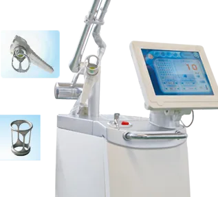
Pmise CF-03
Three output modes: CW, UltraPulse and Fractional.The COHERENT RF-excited metal laser system enables the output quality …
View details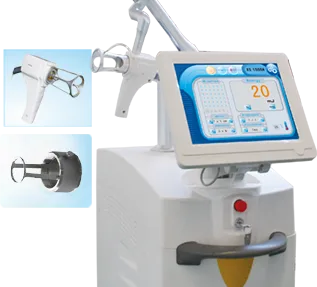
Pmise CF-01
Three output modes: CW, UltraPulse and Fractional.The COHERENT RF-excited metal laser system enables the output quality …
View details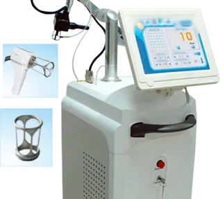
Pmise CF-05
Max. laser power 30W,40W and 50W for optional. Cost-effective with high quality, most competitive for doctors. Most conv…
View details