Pigment Treatment
Pigmented Nevus Treatment: Laser Selection, Protocol and Aftercare
A pigmented nevus (pigment nevus, or melanocytic naevus) is a benign nest of melanocytes in the skin, and pigmented nevus treatment with lasers splits along one line: Q-switched nanosecond pulses for flat superficial pigment, ablative vaporization for raised lesions. Diagnosis comes first, every time.
This is educational material for clinics and distributors comparing equipment, not medical advice. No spec sheet replaces assessment by a clinician.
What a pigmented nevus actually is
Melanocytes migrate from the neural crest during development. Sometimes they bunch up instead of spreading evenly, and that clump is the mole your patient wants gone. Three types matter, and the type picks the machine.
- Junctional nevus. Flat or barely raised, brown through brown-green to black, smooth and hairless. Nests sit at the dermo-epidermal junction, so pigment is shallow.
- Compound (hybrid) nevus. Looks similar but stands higher off the skin, with nests straddling the junction into the dermis.
- Intradermal nevus. Dome-shaped or papillary, sometimes on a stalk, often with hairs through it. Cells lie buried in dermal collagen, little surface melanin.
Sizes run from a few millimetres to a few centimetres, and none of them clear on their own.
Diagnosis before device
Here is the part no brochure should skip. A laser destroys the lesion, and with it the tissue a pathologist needs to rule out melanoma. Treat first and you can bury a cancer under a healed scar. The American Academy of Dermatology's ABCDE checklist is the standard screen. Know it cold.
- Asymmetry: one half doesn't match the other.
- Border: irregular, scalloped or poorly defined edges.
- Color: shades varying across the lesion, or patches of white, red, blue.
- Diameter: melanomas are typically over 6 mm, though smaller ones occur.
- Evolving: changing in size, shape or colour, or unlike the patient's other moles.
Delker and colleagues reviewed melanomas found in skin previously treated by laser (Journal of Dermatology, 2017). In most cases no histological assessment had been carried out beforehand. Their conclusion was blunt: laser therapy of pigmented lesions should be avoided, or performed only after prior histology.
The rule stays simple. Anything irregular, changing, bleeding, itching or new goes to dermoscopy and biopsy, not a handpiece.
How lasers treat pigmented nevi
Photoacoustic fragmentation. A Q-switched Nd:YAG fires nanosecond pulses at 1064 nm and 532 nm. Melanin absorbs that energy faster than it sheds heat, the particle fractures, and phagocytes clear the debris over following weeks. Anderson and Parrish set out the principle in 1983: pick a wavelength the target absorbs, keep the pulse shorter than its thermal relaxation time, and damage stays put. The 1064 nm beam reaches deeper blue-black pigment; 532 nm peaks nearer the surface. This is the route for junctional lesions, where the problem is pigment, not bulk.
Ablative vaporization. A CO2 laser emits at 10600 nm, a wavelength water absorbs avidly. Melanin is irrelevant here. Tissue boils away in layers, which suits a compound or intradermal nevus: you're removing a mound of cells, not scattered pigment. Local anaesthetic is standard, ultra-pulse the usual mode.
Now the caveat. Vaporization leaves nothing behind: no specimen, no slide, no second opinion, even for a lesion that looked ordinary. Ablation fits only where a clinician has examined the lesion and judged it benign. Excision, or shave excision, with tissue sent for histopathologic examination is the alternative. Prefer it when:
- the diagnosis is uncertain, clinically or on dermoscopy;
- dermoscopy shows atypical features, or the patient reports a change;
- risk factors exist: personal or family melanoma history, many nevi, heavy sun damage;
- local regulation or clinic policy requires histology for melanocytic lesion removal.
Both routes share one principle from our engineering archive: thorough elimination of the nevus cells is the key to success. Leave cells behind and it returns.
Treatment protocol and what to expect
Protocols are individual. Depth, diameter, site and Fitzpatrick type move the plan, so treat the table as orientation.
| What you're weighing | Flat, superficial lesion | Raised, cellular lesion |
|---|---|---|
| Device | Q-switched Nd:YAG, 1064 and 532 nm | Ultra-pulse CO2, 10600 nm |
| Specimen for pathology | None | None; excision preserves it |
| Sessions | Usually several, weeks apart | Often one, occasional touch-up |
Talk the patient through this first. Shallow pigment fades step by step over weeks, under topical anaesthetic at most. A raised lesion goes in one sitting under local infiltration, but leaves a wound. Test spots make sense in Fitzpatrick IV to VI.
Who should not be treated
Our manuals carry explicit exclusion lists. Most complications trace back to a patient who should never have been treated.
- Broken skin, active herpes, inflammation or infection at the site. Treating through it wrecks healing.
- Malignant or suspicious lesions nearby. Dermatologist, not handpiece.
- Recent retinoid use, isotretinoin included. Healing is altered, scar risk climbs.
- Keloid or hypertrophic scarring history. Manuals advise against a scarring diathesis.
- Cardiac pacemaker or implanted defibrillator. A stated Q-switched contraindication.
- Severe diabetes, hypertension, heart disease, epilepsy, systemic illness. Poor healing, added risk.
- Pregnancy. Precautionary; none of this is urgent.
- Recent vascular surgery or resurfacing there. Let the skin settle first.
- Recent tanning or photosensitising medication. Both raise burn and dyschromia risk.
- Unrealistic expectations, or refusal to follow sun protection. Listed outright in one manual.
Laser safety in the treatment room
- Eyewear matched to the wavelength. Our Q-switched manual specifies glasses covering 200 to 1080 nm for operator and patient; 532 nm green light is a direct eye hazard. CO2 at 10600 nm needs eyewear rated for it, and Nd:YAG goggles won't do.
- Plume evacuation during CO2 ablation. Vaporized tissue makes surgical smoke. Run an evacuator at the site, plus respiratory protection.
- Control the room. No reflective surfaces, no flammable material, metal off the patient, sign at the door, no traffic while firing.
- Trained operators only. Our manuals restrict operation to trained professionals; credentialing follows local regulation.
More in our laser safety essentials for clinics.
Recommended Pmise equipment
For superficial pigment, a dual-wavelength Q-switched Nd:YAG laser from the QN series covers the colours you meet in practice. Its manual in our engineering archive specifies 1064 nm and 532 nm output, spot diameter adjustable from 1 to 7 mm, single pulse energy up to 800 mJ at 1064 nm and up to 400 mJ at 532 nm, pulse width of approximately 6 ns, and repetition rate from 1 to 10 Hz. Short pulses matter more than raw power.
Underpowered platforms are a false economy. Our clinical archive advises against systems below roughly 200 mJ single pulse for dermal melanocytic lesions: weak clearance, longer courses, more pain, more scarring.
For raised compound and intradermal lesions, an ultra-pulse CO2 laser from the CF series gives layer-by-layer removal. Our CO2 manual lists 10600 nm output, ultra-pulse mode with random scanning, a 0.25 mm focused spot, pulse energy in 2 mJ steps up to 200 mJ, and working power from 1 W to 30 W in 1 W steps. Mixed caseloads need both.
Four commercial questions decide whether a platform is buyable:
- Certification. Ask for the current certification file and confirm what applies where you sell.
- Operator training. Installation, parameter training, written protocols: what actually ships?
- Warranty and after-sales service. Term in writing, plus turnaround on cavity and handpiece repair.
- Consumables and spares. Parts list, lead times, pricing.
Want the QN and CF specification sheets, or a clinical application call about your case mix? Talk to our engineering team. For wavelength and endpoint logic, read our guide to treating pigmented lesions with Q-switched lasers, or see Nevus of Ota for deeper dermal pigment.
Aftercare, risks and recurrence
- Keep the site clean and dry. After CO2 ablation, no water on the wound for a few days.
- No picking. Crusts should separate on their own; forcing them off invites scarring.
- Strict sun protection through healing and for months after. Ultraviolet drives pigment back.
- Counsel on post-inflammatory hyperpigmentation. Our archive lists pigment change, persistent erythema and scarring as occasional complications after CO2 treatment of raised nevi; darker skin types carry more risk.
- Be honest about recurrence: deep lesions leave more room for residual cells.
- Re-examine at follow-up. If pigment returns irregularly, refer rather than firing again.
Frequently Asked Questions
Can any mole be removed with a laser?
No, and treating it that way is how people get hurt. Flat junctional pigment suits a Q-switched laser; raised lesions need ablation. Both destroy the lesion, so nothing survives for pathology. Ablation belongs only to lesions a clinician has judged benign. Where doubt exists, excision with histopathology is correct.
How many sessions does a pigmented nevus need?
It depends on the lesion, so avoid promising a number. Superficial pigment treated with a Q-switched laser usually lightens gradually over several sessions weeks apart, which gives the body time to clear fragmented pigment. A raised lesion removed ablatively is often one session. Depth, size and skin type shift the count.
Will the nevus grow back?
It can, and the reason is simple: nevus cells left in place. Complete removal is the goal for exactly that reason. Deep intradermal lesions carry more recurrence risk than shallow ones. If pigment reappears with an uneven border or mixed colour, refer for reassessment instead of re-treating on autopilot.
What about scarring and pigment change?
Both are real risks with any energy device. Ablation creates a wound, and poor aftercare turns wounds into scars. Either approach can trigger post-inflammatory hyperpigmentation, and that risk climbs in darker Fitzpatrick types. Conservative parameters, a test spot and disciplined sun protection help.
Evaluating a Q-switched or CO2 platform for pigmented lesion work? Request the specification sheets or book a clinical application consultation, and we'll walk through parameters, training and service.
Recommended Equipment
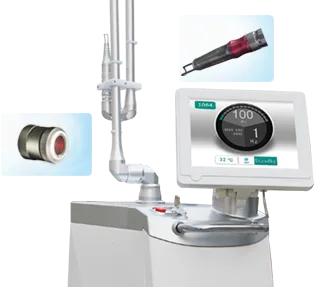
Pmise QE-01
Dual wavelengths (1064nm & 532nm) are convenient to adjust by touching LCD. Multi-spot size (1-7mm) is ideal for a wide…
View details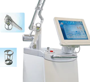
Pmise CF-03
Three output modes: CW, UltraPulse and Fractional.The COHERENT RF-excited metal laser system enables the output quality …
View details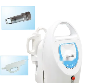
Pmise QN-05
Patented technology of single lamp and dual rods doubles energy output (400mj). Shorter pulse width (6-8ns) than ordina…
View details