
Pigment Treatment
Nevus Laser Treatment: Lesion Types, Biopsy-First Protocol and Device Selection
A nevus is a benign cluster of pigment cells parked in the skin, and nevus laser treatment uses Q-switched or ablative light to shatter or vaporise that pigment. On the right lesion it works well. On the wrong one it destroys the evidence a pathologist needed.
This page is for clinics, dermatologists and distributors comparing equipment. It is educational, not medical advice, and no machine replaces clinical assessment.
What a nevus is, and why the type decides the tool
Melanocytes migrate toward the epidermis before birth. Sometimes they bunch up instead of spreading evenly. You get a mole, medically a melanocytic nevus, and the depth those cells settled at decides whether your laser can clear it. The clinical atlas in our engineering archive sorts pigment nevi three ways.
- Junctional nevus. Nests sit at the dermal-epidermal junction. Flat, brown to brown-black, smooth, usually hairless. Shallow pigment.
- Compound nevus. Nests straddle the junction and the dermis. Picture a junctional lesion grown upward out of the skin.
- Intradermal nevus. Cells sit deep, wrapped in collagen, often carrying little melanin. Skin-coloured or pink as often as dark, usually with a coarse hair pushing through.
A fourth pattern sits in our archive: congenital light brown patch-like pigmentation carrying darker brown papules, known as nevus spilus. It appears in infancy, spreads through adolescence, then settles. Blue nevus and congenital melanocytic nevus round out the list, the giant forms needing surveillance rather than a handpiece.
Biopsy first, laser second
Clinics skip this part. It is also the part that ends careers.
Light destroys tissue. It does not hand anybody a specimen. Treat an early melanoma with a Q-switched pulse and you wipe the surface clean while whatever sits underneath keeps growing. Delker and colleagues documented precisely this in the Journal of Dermatology in 2017, reviewing patients who developed melanoma in skin previously lasered. In most of those cases nobody had taken histology first.
So the rule is short. Anything atypical goes to a dermatologist, anything questionable gets excised and sent to pathology, and the laser waits for the report. The American Academy of Dermatology publishes the ABCDE shorthand: asymmetry, irregular border, uneven colour, diameter above roughly 6mm, evolution over time. Itching, bleeding, fast change? Pathologist, not laser.
Biopsy first is not a legal formality. It is the difference between a cosmetic result and a missed cancer.
How laser and light clear nevus pigment
Two mechanisms. Two very different machines.
The first is selective photothermolysis, set out by Anderson and Parrish in Science in 1983. Deliver a pulse shorter than the target's thermal relaxation time, at a wavelength melanin absorbs well, and the pigment particle fractures before heat leaks into normal skin. That is the Q-switched Nd:YAG. Nanosecond pulses at 1064nm reach deep dermal pigment; the 532nm beam suits shallower targets but gets soaked up by epidermal melanin, so it risks more in darker skin.
Ablative vaporisation is the second route. A CO2 laser at 10600nm is absorbed by tissue water, not melanin, so it removes the lesion layer by layer, reaching nests a Q-switched pulse never will. Our archive is blunt about the pairing: junctional lesions take Q-switched Nd:YAG, compound and intradermal lesions need ultra-pulse CO2 with local anaesthetic.
Leave cells behind and you have created a second problem, not just a cosmetic disappointment. Pigment returning inside a treated site is a recurrent nevus. Kornberg and Ackerman named that pattern pseudomelanoma in 1975 for good reason: down the microscope it borrows melanoma's features, cytological atypia and pagetoid spread among them, and a pathologist who does not know a laser was there can read it wrongly. Repigmentation is a dermatology referral, never a repeat session. Hand over the before photographs and write the prior laser treatment on the pathology form.
Laser or excision? Matching the lesion to the method
Excision wins on two counts. It produces a specimen for histology, and it clears the full depth in one pass. You pay with a linear scar. Laser trades that scar for a course of sessions and, on raised lesions, some risk of a dent.
| Lesion | Reasonable approach | Notes |
|---|---|---|
| Flat junctional nevus, benign on assessment | Q-switched Nd:YAG, 1064nm or 532nm | Smaller spot, higher fluence; several sessions |
| Compound or intradermal nevus, raised | Ablative CO2 vaporisation | Local anaesthesia; often single-stage |
| Nevus spilus background patch | Q-switched Nd:YAG, energy headroom at a wide spot | Darker papules may need ablative work |
| Any atypical or changing lesion | Excision and histology | No laser until pathology reports |
| Repigmentation in an old treatment site | Dermatology assessment | Disclose the prior laser treatment to the pathologist |
For the wider picture, read our overview of pigmented lesions and Q-switched laser selection and the dedicated pigmented nevus treatment page.
Protocol, energy and what to expect
Millijoules on a brochure tell you nothing alone. Effect on melanocytic pigment tracks fluence, in joules per square centimetre, and that depends on the spot diameter your operator dials in. Run the arithmetic on one 400mJ pulse:
- Through a 2mm spot it lands at roughly 13 J/cm2.
- Through a 4mm spot, about 3.2 J/cm2.
- Open up to 8mm and you are near 0.8 J/cm2, sixteen times weaker than the 2mm figure.
Same pulse energy. Wildly different treatments. Work to a fluence and a visible endpoint, never to a number copied off a spec sheet.
Where does archive guidance fit? Our engineering archive advises against Q-switched platforms delivering below roughly 200mJ per pulse on melanocytic lesions, citing weak effect, longer courses, more pain and more scarring, and treats around 400mJ single-pulse as the point where an operator can widen the spot and still hold useful fluence. Both figures are in-house guidance for that device class, not an industry threshold. Read them as a question about energy headroom, never a clinical dose.
- Sessions. Flat pigment usually needs several passes weeks to months apart. Deeper lesions take longer. Raised lesions handled ablatively often clear in one or two.
- Endpoint. Immediate whitening on Q-switched passes. Controlled removal without charring on ablative work.
- Downtime. Redness and crusting for days after ablative work. Q-switched passes are gentler but still crust, and that crust must be left alone.
- Recurrence. Pigment can come back, and patients deserve to hear that before session one. Incomplete removal can leave a recurrent nevus whose histology mimics melanoma closely enough to confuse a later biopsy, so a repigmented site goes to a dermatologist with the treatment history, not back under the handpiece.
Recommended Pmise equipment
Most clinics here end up owning two platforms.
For flat, melanin-rich targets, our Q-switched Nd:YAG laser range covers the ground, from compact QN-series units up to the electro-optic models. Per our device manuals, the QE-01 runs a 6ns pulse, roughly 800mJ at 1064nm and 420mJ at 532nm, and a flat-top beam that spreads energy evenly instead of spiking in the centre. Spot size adjusts from 1 to 8mm, which is the number worth reading: at 8mm that 1064nm pulse lands near 1.6 J/cm2, while pulling the spot to 3mm pushes it into double figures.
Raised lesions want the ultra-pulse CO2 platform. The CF-01 runs 10600nm from an RF-excited tube at 30W, with continuous, ultra-pulse and fractional modes, pulse energy from 10 to 200mJ and a focal spot from 80 to 2000 micrometres. Fine spot control lets an operator take a papule down cleanly rather than gouging a crater.
Aftercare and precautions
- Leave crusts alone until they separate. Picking causes scarring and post-inflammatory hyperpigmentation.
- Strict sun protection for weeks. Freshly treated skin pigments readily.
- Fitzpatrick IV and above need lower fluences, wider spots, longer intervals. Post-inflammatory hyperpigmentation is the commonest complication in darker skin and can take months to fade.
- Screen contraindications: photosensitising drugs, active herpes, keloid history, uncontrolled diabetes, pregnancy.
- Photograph everything before you treat. If pigment reappears, that record tells a dermatologist whether it is residual nevus or new disease.
Frequently Asked Questions
Can a laser remove a mole permanently?
Sometimes. Permanent clearance depends on eliminating every nevus cell, and cells deep in the dermis are hard to reach without ablation. Raised lesions treated with CO2 often clear in one stage. Flat lesions may fade over several sessions, then partly return. Incomplete removal can leave a recurrent nevus that imitates melanoma under the microscope and makes any later biopsy hard to read, so a repigmented site goes to a dermatologist with the laser history disclosed.
Why does a biopsy matter if the mole looks harmless?
Because laser destroys the tissue a pathologist would have examined. Published case series document melanomas found in skin previously lasered, generally where no histology was taken first. A trained eye is good. It is not infallible. Anything asymmetric, irregularly bordered, multi-coloured, over about 6mm or visibly changing belongs in a specimen pot.
How should we compare single-pulse energy between machines?
Ask for pulse energy and spot range together, then convert to fluence yourself. A millijoule figure with no spot diameter beside it is not a specification, it is marketing. You want enough energy at the handpiece to hold a useful fluence at the widest spot your protocols call for, plus a stable pulse width and flat-top profile.
Next step: specifications, parameters and distributor enquiries
- Clinics and dermatology practices: request the full Q-switched Nd:YAG and ultra-pulse CO2 specification sheets, including pulse width, spot range and measured energy at the handpiece, then book a parameter consultation with our engineering team.
- Distributors and importers: ask for the distributor pack, covering regional pricing, certification files, operator training material and the biopsy-first protocol you will need to teach your accounts.
Tell us which lesions and skin phototypes you treat most, and we will map them to a configuration rather than push a catalogue.
Recommended Equipment
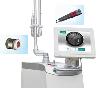
Pmise QE-01
Dual wavelengths (1064nm & 532nm) are convenient to adjust by touching LCD. Multi-spot size (1-7mm) is ideal for a wide…
View details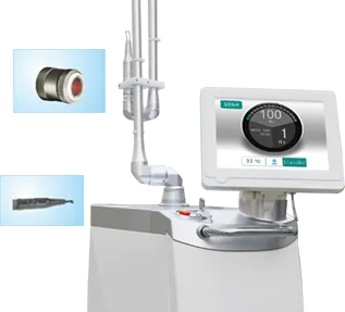
Pmise QE-02
Dual wavelengths (1064nm & 532nm) are convenient to adjust by touching LCD. Multi-spot size (1-7mm) is ideal for a wide…
View details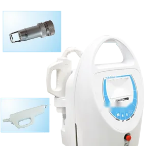
Pmise QN-05
Patented technology of single lamp and dual rods doubles energy output (400mj). Shorter pulse width (6-8ns) than ordina…
View details