Pick the wavelength the blood absorbs, then pick the box around it. Hemangioma treatment lives or dies on that match. Red and superficial? Use a vascular laser or filtered light tuned close to where oxyhaemoglobin absorbs. Bulky, bluish, deeper? Go longer, out to 1064 nm, trading absorption for reach. A proliferating infantile hemangioma is a different conversation, and it starts with a paediatrician.
Is it actually a hemangioma?
Most vascular mistakes happen before the laser fires. "Hemangioma" gets stuck on anything red and raised, yet the label covers conditions that behave nothing alike. Infantile hemangiomas appear in the first weeks of life, grow, then involute. Cherry angiomas in adults stay small and stable. Our clinical training atlas is clear: many infantile lesions involute unaided, and treatment is reserved for cases where function is at risk.
Colour and depth decide the rest. Bright red near the surface absorbs visible light well. Bluish and bulky means reaching into the dermis, and only a longer wavelength gets there.
| Lesion | What you see | Depth | First move |
|---|---|---|---|
| Infantile hemangioma, proliferating | Raised red "strawberry" mark, infants | Superficial to deep | Specialist review first |
| Cherry angioma | Small bright red dome, adults | Superficial | Vascular laser or high-frequency coagulation |
| Telangiectasia, spider veins | Fine red threads, face or legs | Papillary dermis | Vascular laser, IPL, or a point tool |
| Port-wine stain | Flat pink to purple patch, from birth | Dermal | Pulsed dye laser, several sessions |
A port-wine stain is a capillary malformation, not a hemangioma. Depth decides your wavelength; the badge on the panel doesn't.
Why oxyhaemoglobin decides your wavelength
Vascular lasers run on selective photothermolysis, the principle Anderson and Parrish published in Science in 1983. Choose a wavelength the target absorbs well. Deliver it in a pulse short enough to trap the heat inside. Light lands in the blood, becomes heat, and the vessel wall is damaged while the skin around it is largely spared.
The chromophore is oxyhaemoglobin inside the vessel. The IADVL standard guidelines of care for lasers on vascular lesions put its major absorption peaks at 418, 542 and 577 nm, with optimal absorption between 577 and 600 nm, and our internal notes list the same peaks.
One detail gets skipped in most sales conversations. Heated haemoglobin oxidises into methaemoglobin, and our technical archive records that methaemoglobin absorbs 1064 nm around thirteen times more strongly than deoxyhaemoglobin, three times more than oxyhaemoglobin. Part of why a long-pulse 1064 nm device still does serious vascular lesion work from nowhere near the visible peaks.
Which wavelength for which vessel?
No wavelength wins everywhere. There's a best match for a given depth and colour, and the trade-off is absorption against reach.
| Technology | Wavelength | Best fit | Skin type suitability | Trade-off |
|---|---|---|---|---|
| Pulsed dye laser | 585 / 595 nm | Superficial redness, port-wine stains, thin hemangiomas | Most predictable on Fitzpatrick I-III; on IV-VI cut energy, lengthen the pulse, cool hard | Transient purpura possible |
| KTP | 532 nm | Fine facial telangiectasia, cherry angiomas | Least forgiving on darker skin, since epidermal melanin absorbs strongly here | Limited depth, competes with melanin |
| Long-pulse Nd:YAG | 1064 nm | Deeper or larger vessels, leg veins | Usual first choice for Fitzpatrick IV-VI, and for deep vessels on any skin | Weaker absorption, careful fluence control |
| IPL and E-light | Broadband, filtered | Diffuse background redness and flushing | Move to a longer cut-off filter on darker skin to protect the epidermis | Less selective than a single-wavelength laser |
| High-frequency coagulation | Not light-based | Pinpoint vessels, small cherry angiomas | Not wavelength dependent, though darker skin marks more readily at treated points | Vessel by vessel, operator dependent |
Skin type changes the answer, not only the settings
Melanin sits in the epidermis and absorbs strongly across the visible range, so at 532, 585 and 595 nm the surface competes for light you meant for the blood. On Fitzpatrick IV-VI skin that competition raises the risk of epidermal burns, blistering and post-inflammatory hyperpigmentation. Our training material lists pigment change as a recognised sequel of visible-wavelength vascular work, managed with strict sun protection and longer gaps between sessions, and sets out the mitigations for darker skin: longer wavelength, longer pulse width, reduced output energy, active epidermal cooling, and on filtered systems a longer cut-off filter. Which is exactly why 1064 nm becomes the workhorse in Middle Eastern, African and South East Asian clinics. Melanin barely absorbs it, and it reaches deeper vessels anyway. Selling into those markets? Quote the Nd:YAG platform first. Whatever the wavelength, test spot on Fitzpatrick IV-VI skin and wait longer than usual before committing to the whole lesion.
Pulse duration deserves as much attention as the nanometre figure. Two devices can both say 1064 nm and behave nothing alike, the point of our note on long-pulse versus Q-switched Nd:YAG. Want the physics? Thermal relaxation time covers it, and our overview of vascular conditions treated with Nd:YAG shows where 1064 nm earns its keep.
What equipment do clinics actually buy?
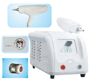
Hardly anyone builds a vascular service on one machine. A pulsed dye or KTP system takes the superficial work. A long-pulse Nd:YAG adds depth and doubles as a hair removal platform, which is usually what makes the purchase pay. Our device manuals name a long-pulse 1064 nm system with adjustable pulse count and pulse width plus contact cooling as first choice for hemangioma, with a contact-cooled 10 mm by 10 mm handle for larger lesions.
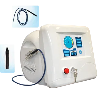
For the smallest lesions a laser is overkill. High-frequency coagulation is faster and cheaper to run. Our device documentation describes a high-frequency approach that thermally coagulates the haemoglobin and endothelial cells of a dilated capillary through a tip roughly 0.01 mm across, so the epidermis is largely spared. The units run at 13.56 MHz, continuous or pulsed. Tolerance is generally good, with most patients reporting brief stinging, and topical anaesthetic stays optional for longer sittings. That is how the Pmise VF-05 in our high-frequency range for vessel diseases works. A salon clearing cherry angiomas between facials gets more from a fine tip than a large spot.
Then the boring part, which decides running costs and compliance risk.
- Regulatory clearance. Which certificates name the exact model you're buying: CE, FDA 510(k), local registration? A sister model's paperwork won't clear customs.
- Wavelengths and spot sizes. Can the platform take a second handpiece later? Confirm which spot sizes ship in the box.
- Pulse width and pulse number. This decides whether you heat the vessel or the tissue around it. Ask for the real adjustable range, not "fully variable".
- Cooling method. Cryogen spray, contact sapphire, or cold air. Our training material ranks them in that order for vascular work, and warns that any epidermal cooling means raising output energy.
- Consumables. Dye cells, flashlamps, tips, filters. Get replacement price and expected life, then work out cost per treatment. That beats comparing sticker prices.
- Training and protocols. Parameter guidance by lesion type and Fitzpatrick skin type, and cover for new operators.
- Warranty, spares and installation. Exclusions, service location, dispatch time for critical parts, plus voltage and mains frequency for your market.
- Lead time and MOQ. Minimum order quantity, production lead time, and whether local labelling is supported.
Not sure what fits the lesions you actually see? Our hemangioma solution page maps lesions to devices. Send your case mix and usual skin types via the contact page and we'll come back with a wavelength and cooling recommendation plus the model comparison sheet.
When do you refer instead of treat?
Refer when it's a proliferating infantile hemangioma. The American Academy of Pediatrics Clinical Practice Guideline for the Management of Infantile Hemangiomas, published in Pediatrics in 2019, makes oral propranolol the drug of choice when systemic treatment is indicated. Laser isn't a replacement there. Its role comes later, on residual skin change.
Send these onward the same day, not into your booking system:
- Lesions near the eye, lip, nose tip or airway, where growth threatens function.
- Ulceration, bleeding, or rapid enlargement.
- Multiple lesions, or a large segmental facial lesion that may signal a syndrome.
- Any doubt about whether it's a hemangioma at all.
Adult lesions that are safe to treat still demand discipline. Assess Fitzpatrick type, use proper eyewear, test spot first, and tell patients honestly that several sessions are usually needed and results vary with lesion and skin type. The European Society for Laser Dermatology guidelines on vascular lasers and intense pulsed light, published in JEADV in 2015, are a sensible backbone for your protocol.
Cooling, and the rest of a safe workflow
Cooling separates good operators from average ones, and it isn't one setting for every case. Our training material is blunt: fine telangiectasia shouldn't be cooled aggressively, since those vessels sit near the epidermis and over-protecting the surface leaves treatment ineffective. Hemangiomas invert that. They need higher energy density, so epidermal cooling is required, and our manuals recommend contact sapphire cooling at around minus 4 degrees with the larger handles. On darker skin cooling stops being a comfort feature, and output energy comes down rather than up.
- Confirm the diagnosis; screen out infantile and high-risk lesions.
- Photograph the lesion and record Fitzpatrick skin type.
- Match wavelength and platform to lesion depth, colour and skin type.
- Test spot, then read the tissue response before treating the area.
- Fit eye protection, set cooling for the lesion, start conservative.
- Give written aftercare and space sessions several weeks apart.
- Review at every visit rather than chasing clearance in one pass.
Frequently Asked Questions
Can a laser remove a hemangioma in one session?
Rarely, and only for the smallest superficial lesions. A tiny cherry angioma or fine telangiectasia may clear after one treatment. Most vascular lesions need a course spaced several weeks apart. Marketing that promises single-session clearance only sets patients up to be disappointed.
Which wavelength suits darker skin?
Start with 1064 nm. Melanin absorbs it weakly, so the epidermis competes less and the risk of burns and post-inflammatory pigment change falls compared with 532, 585 or 595 nm. On Fitzpatrick IV-VI skin, use longer pulse widths, lower output energy and active cooling. Test spot first, then leave a longer gap before treating the full lesion.
Should I ever treat an infant's hemangioma with a laser?
Not as a first step, and not without a paediatric specialist leading the case. Under the AAP guideline, oral propranolol is the drug of choice when systemic treatment is needed, with laser mostly reserved for residual skin changes once the lesion settles. The safest role for an aesthetic clinic is to spot these cases and refer them on.
Published 22 April 2026, reviewed July 2026. Compiled by the Pmise Technical Team, our product engineering and clinical applications group, and checked against the sources listed here and our device manuals. This is device selection guidance for licensed practitioners and distributors, not medical advice. Diagnosis and parameters stay with the treating practitioner, and infantile cases must be led by a paediatrician.